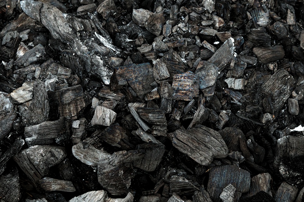
Rosnące zainteresowanie alternatywnymi źródłami ogrzewania sprawia, że hurtowa sprzedaż brykietu staje się ważnym elementem rynku paliw opałowych. Brykiet jest ceniony za wygodę użytkowania, wysoką wartość opałową oraz możliwość efektywnego przechowywania. […]

Mieszkania do wynajęcia – elastyczna alternatywa dla kredytu
Mieszkania do wynajęcia Mogilno to rozwiązanie, które zyskuje na popularności wśród osób ceniących elastyczność i brak długoterminowych zobowiązań. W dynamicznie zmieniającej się rzeczywistości wynajem staje się realną opcją zarówno dla […]
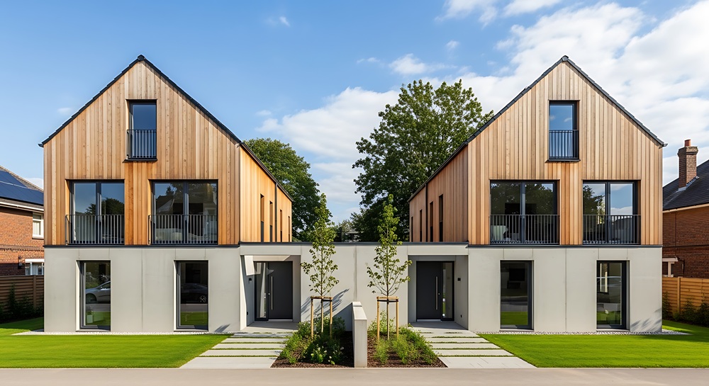
Bliźniaki – funkcjonalne rozwiązanie dla nowoczesnych rodzin
Bliźniaki (zabudowa bliźniacza) to coraz częściej wybierana forma zabudowy mieszkaniowej, która łączy w sobie komfort domu jednorodzinnego z optymalizacją kosztów budowy i utrzymania. To rozwiązanie szczególnie atrakcyjne dla osób szukających […]
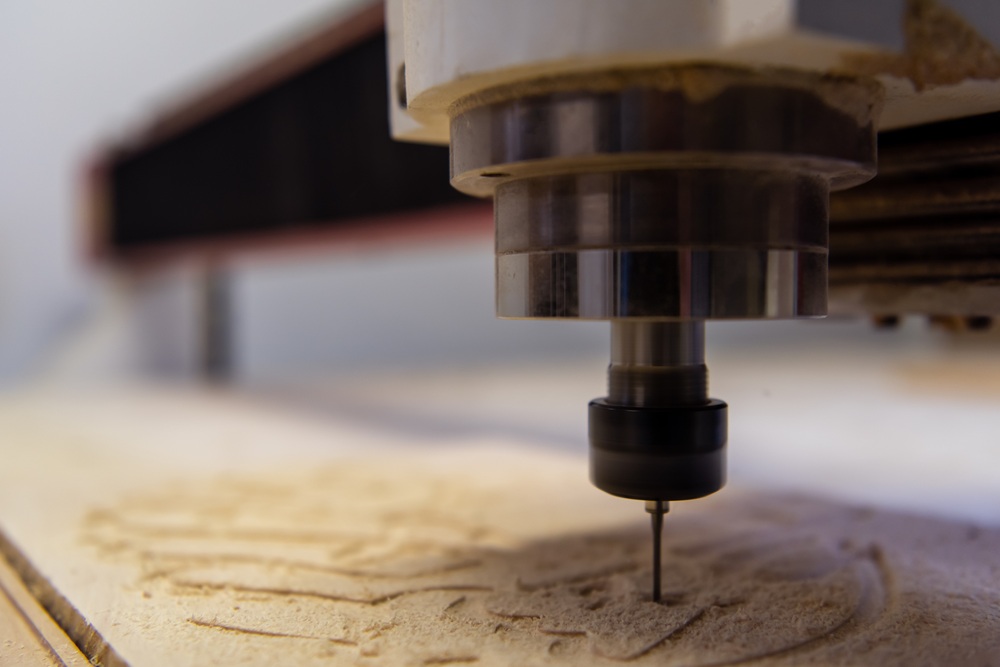
Frezowanie CNC – precyzyjne usługi CNC dla drewna i innych materiałów
Frezowanie CNC Bydgoszcz to jedna z najnowocześniejszych technologii obróbki materiałów, która pozwala na osiągnięcie wyjątkowej precyzji i powtarzalności w produkcji. Wykorzystując sterowanie komputerowe, maszyny CNC mogą wykonywać skomplikowane kształty w […]